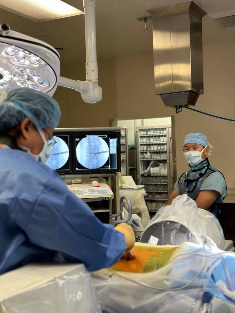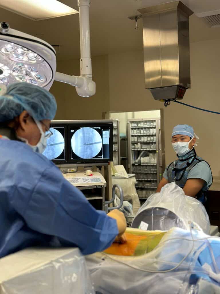
Minimally invasive spine surgery (MIS) has revolutionized spine surgery by offering patients quicker recoveries, reduced pain, and minimal scarring compared to traditional open surgeries. Central to success of MIS is the use of navigation technology, which enhances precision and safety. In this blog, we explore the crucial role that navigation plays in minimally invasive spine surgery and how it benefits both patients and surgeons.
Introduction to MIS
Minimally invasive spine surgery aims to achieve the same surgical objectives as traditional open surgery but through smaller incisions. This approach minimizes muscle and tissue damage, leading to faster recovery times and less postoperative pain. Techniques such as tubular retractors, endoscopic cameras, and advanced imaging technologies are employed to access the spine with minimal disruption.
Importance of Navigation in Spine Surgery
Navigation systems in spine surgery are like GPS for surgeons. They provide real-time, three-dimensional imaging of the patient’s anatomy, allowing surgeons to plan and execute procedures accurately. These systems integrate preoperative CT or MRI scans with intraoperative images, creating a dynamic roadmap that guides the surgeon throughout the operation.

Key Benefits of Navigation in Minimally Invasive Spine Surgery
1. Enhanced Precision
Navigation technology allows for precise localization of spinal anatomy and pathology. This precision is particularly critical when dealing with delicate structures such as nerves and blood vessels. The ability to navigate accurately ensures that surgical instruments are placed with millimeter-level accuracy, reducing the risk of complications and improving outcomes.
2. Reduced Radiation Exposure
Traditional spine surgeries often rely on fluoroscopy, which exposes patients and surgical staff to significant radiation. Navigation systems minimize the need for continuous fluoroscopic guidance by providing accurate real-time imaging, thereby reducing overall radiation exposure.
3. Improved Surgical Outcomes
Studies have shown that navigation-assisted MIS results in higher accuracy in pedicle screw placement, reduced surgical time, and lower rates of complications compared to non-navigated procedures. These factors contribute to improved patient outcomes and satisfaction.
4. Shorter Recovery Times
By minimizing tissue damage and ensuring precise surgical interventions, navigation in MIS leads to shorter hospital stays and quicker return to daily activities for patients. The reduced postoperative pain and faster recovery times are significant benefits that enhance the overall patient experience.
How Navigation Systems Work
Preoperative Planning
Before surgery, detailed imaging studies such as CT or MRI scans are performed to map the patient’s spinal anatomy. These images are then uploaded into the navigation system, where they are used to create a comprehensive surgical plan. Surgeons can simulate the procedure, plan the optimal surgical approach, and anticipate potential challenges.
Intraoperative Navigation
During the surgery, the navigation system continuously updates the surgeon with real-time images. A reference frame is attached to the patient to correlate the preoperative images with the actual anatomical position. Instruments equipped with tracking devices are used, and the navigation system displays their position relative to the patient’s anatomy on a monitor, guiding the surgeon’s movements with high precision.
Postoperative Verification
After the procedure, navigation systems can be used to verify the accuracy of the surgical intervention. This step ensures that implants are correctly positioned, and any necessary adjustments can be made before concluding the surgery.
Conclusion
Navigation technology has become an indispensable tool in minimally invasive spine surgery, offering enhanced precision, reduced radiation exposure, improved outcomes, and shorter recovery times. As technology continues to advance, the future holds even greater promise for navigation-assisted MIS, ensuring that patients receive the safest and most effective spinal care possible.
FAQs
What is minimally invasive spine surgery (MIS)? Minimally invasive spine surgery (MIS) involves performing spinal procedures through small incisions using specialized instruments and advanced imaging techniques. This approach results in less muscle and tissue damage, reduced postoperative pain, smaller scars, and quicker recovery times compared to traditional open surgery. MIS surgery is used to treat various spinal conditions, including herniated discs, spinal stenosis, and scoliosis.
How does navigation technology improve surgical outcomes? Navigation technology improves surgical outcomes by allowing precise localization of spinal anatomy, reducing the risk of complications, minimizing radiation exposure, and ensuring accurate placement of surgical instruments and implants.
What are the benefits of minimally invasive spine surgery? Benefits of minimally invasive spine surgery include smaller incisions, less tissue damage, reduced postoperative pain, shorter hospital stays, and quicker recovery times compared to traditional open surgery.
How does navigation reduce radiation exposure in spine surgery? Navigation reduces radiation exposure by minimizing the reliance on continuous fluoroscopic guidance, as it provides accurate real-time imaging without the need for constant X-ray use.
Is navigation technology safe for all patients? Yes, navigation technology is safe for most patients undergoing minimally invasive spine surgery. It enhances the safety and accuracy of the procedure, leading to better outcomes. However, surgeons will evaluate each patient’s specific condition and medical history to determine the best approach. Patients with severe spinal deformities or previous spinal surgeries may require additional considerations.
Can all spine surgeons use navigation technology? While navigation technology is widely available, not all spine surgeons are trained in its use. It’s important to choose a surgeon with specific experience and expertise in navigation-assisted minimally invasive spine surgery. Surgeons with extensive training and proficiency in navigation technology can leverage its full potential to enhance surgical outcomes and patient safety.
What is the difference between MIS and traditional open spine surgery? Minimally invasive spine surgery and traditional open spine surgery differ primarily in incision size, tissue disruption, and recovery. MIS uses smaller incisions, causing less muscle and tissue damage, resulting in shorter hospital stays, faster recovery, less postoperative pain, and smaller scars. Advanced imaging technologies guide the surgery with specialized tools, minimizing complications like infection and blood loss. Traditional open surgery requires larger incisions, more tissue dissection, longer recovery times, and greater postoperative pain and scarring, but it is necessary for more complex spinal conditions. The choice between the two depends on the specific condition, patient’s health, and surgeon’s expertise.
What is a tubular retractor? It is designed to gently separate and hold back muscles and tissues, creating a small working channel through which the surgeon can access the spine. The tubular retractor helps minimize tissue damage by avoiding the need for large incisions and extensive muscle dissection. It allows the surgeon to perform precise surgical maneuvers with specialized instruments while maintaining a clear view of the surgical site through the use of a microscope or endoscope.
What is an endoscope? An endoscope is a medical instrument used to visualize the interior of a body cavity or organ. It consists of a long, flexible or rigid tube equipped with a light source and a camera, allowing doctors to see inside the body without making large incisions. The camera transmits images to a monitor, providing a clear and magnified view of the area being examined or treated.
What are the kinds of advanced imaging technologies used in spine surgery? Fluoroscopy, computer-assisted navigation, intraoperative CT, endoscopic surgery, and O-arm imaging system.
Fluoroscopy is a type of medical imaging that shows a continuous X-ray image on a monitor, allowing real-time visualization of the spine during surgery. This technology guides the placement of instruments and implants, ensuring accuracy and reducing the risk of damaging surrounding tissues. By providing live feedback, fluoroscopy helps surgeons make precise adjustments as needed throughout the procedure.
Computer-assisted navigation combines preoperative imaging data with real-time tracking during surgery to provide a 3D model of the patient’s spine. This technology helps surgeons navigate complex spinal anatomy with high precision, improving the accuracy of implant placement and overall surgical outcomes. It enhances the surgeon’s ability to perform intricate procedures with greater confidence and reduced risk.
Intraoperative CT provides detailed cross-sectional images of the spine during surgery. This allows for immediate assessment and adjustment of surgical techniques, ensuring proper alignment and placement of spinal hardware. By offering high-resolution images in real-time, intraoperative CT helps in identifying any issues that may arise during the operation, allowing for prompt corrections.
Endoscopic Surgery Utilizes an endoscope—a flexible or rigid tube with a camera and light source—to provide a direct view of the surgical area. This allows surgeons to perform procedures through small incisions with real-time visualization, minimizing tissue disruption. Endoscopy is especially beneficial in minimally invasive spine surgery, where it reduces postoperative pain and speeds up recovery.
The O-arm imaging system is a mobile, intraoperative imaging device that provides high-quality 2D and 3D images. It enhances surgical accuracy and safety by allowing for real-time assessment and adjustments during spine surgery. The O-arm system is particularly useful for verifying the placement of screws and other implants, ensuring optimal surgical outcomes..
