
Patients always ask me, “What is the difference between X-ray, CT and MRI and why do I need them?” Well, when it comes to spine-related issues, choosing the right imaging method is crucial for an accurate diagnosis and effective treatment. Whether you’re experiencing back pain, neck pain, a herniated disc, or suspect a more serious condition, understanding the differences between CT scans, MRIs, and X-rays can help you and your doctor determine the best treatment approach. Each imaging technique offers unique insights into the structure of the spine and surrounding tissues. In this blog, we’ll explore the key differences between these three imaging options, helping you understand which might be the most appropriate for diagnosing your spine-related condition.
X-Ray
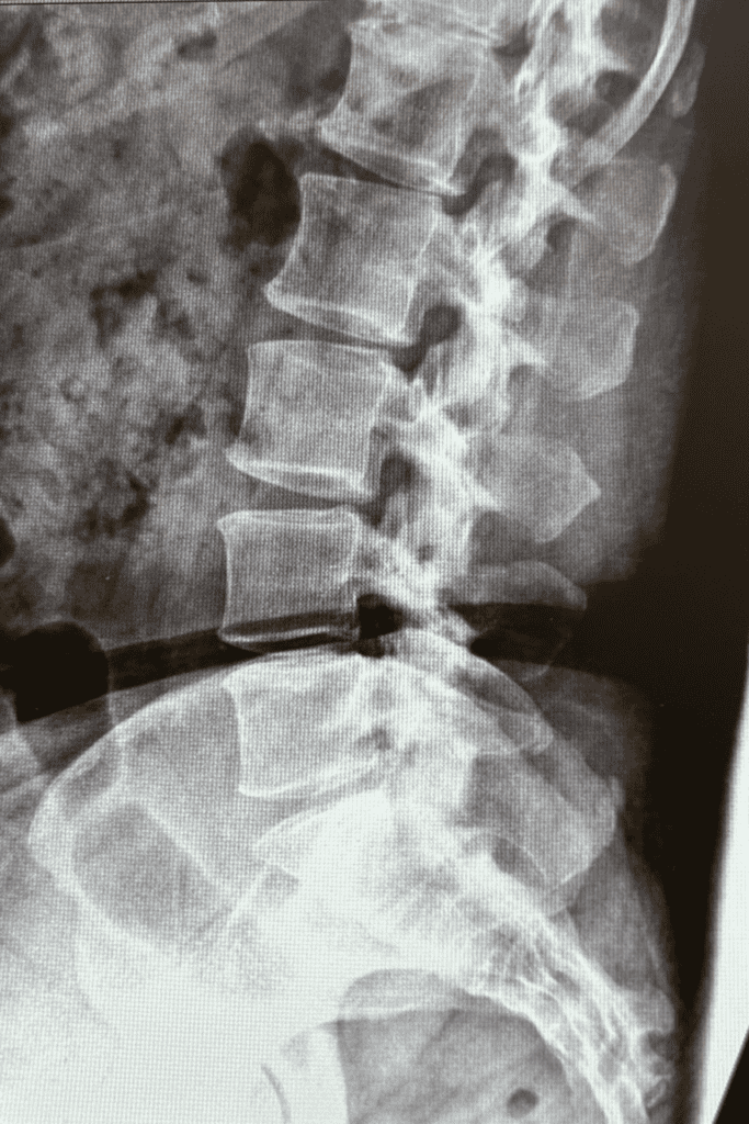
Here are some key points about how X-rays are used in spine imaging:
- X-rays create clear, two-dimensional images of bones, helping diagnose fractures and other bone-related conditions.
- Images can be taken while the patient is standing, allowing doctors to assess the spine under natural weight-bearing conditions.
- X-rays are particularly useful for evaluating the alignment of vertebrae, aiding in the diagnosis of conditions like scoliosis or other misalignments.
- Flexion and extension views can assess spinal stability by showing how the spine moves during bending and extending motions, helping detect issues with movement or stability.
MRI

Here are some key points about how MRIs are used in spine imaging:
- MRIs create detailed 3D images through multi-plane slices, allowing for comprehensive views of the spine from various angles and depths.
- This imaging technique is particularly effective for evaluating soft tissues, including nerves, discs, muscles, tendons, ligaments, and blood vessels.
- MRIs are less effective for examining bones, making them less ideal for bone-specific conditions.
- The imaging process typically requires the patient to lie down to ensure clear and consistent images.
- MRIs are not suitable for individuals with certain implanted devices, such as pacemakers, cochlear implants, or some types of prosthetics, due to the use of strong magnetic fields.
CT Scan
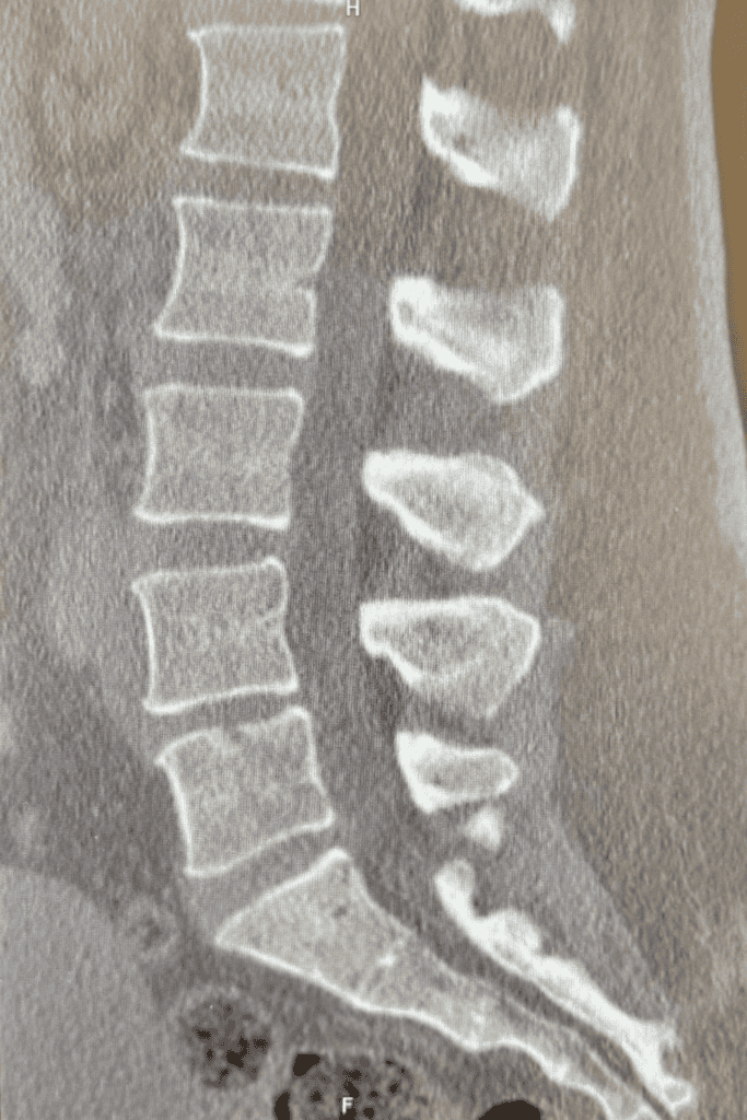
Here are some key points about how CT Scans are used in spine imaging:
- CT scans create detailed 3D images through multi-plane slices, providing comprehensive views of the spine from multiple angles.
- They are particularly effective for evaluating bone structures and are often referred to as the “MRI for bone” due to their detailed bone imaging capabilities.
- CT scans are highly beneficial for pre-operative evaluations, especially in patients with previous surgeries or severe spinal deformities.
In conclusion, selecting the right imaging method—whether it’s an X-ray, MRI, or CT scan—is essential when dealing with spine-related issues. Each technique provides unique insights into different aspects of the spine, from bones and alignment to soft tissues and nerves. By understanding the strengths of each imaging method, you can work with your healthcare provider to make an informed decision that leads to an accurate diagnosis and a targeted treatment plan for your spine condition.
Click here to out this short video on why you need x-rays if you already have an MRI.
FAQs
What is the difference between an X-ray, MRI, and CT scan for spine imaging? X-rays provide two-dimensional images of bones, making them ideal for diagnosing fractures and evaluating alignment. MRIs create detailed 3D images of soft tissues like nerves, discs, and muscles, but are less effective for bones. CT scans, on the other hand, provide 3D images that are excellent for detailed bone evaluation, often referred to as the “MRI for bone.”
Are X-rays enough to diagnose all spine-related conditions? X-rays are great for assessing bone alignment, fractures, and stability, but they may not show detailed soft tissue problems like nerve damage or herniated discs. In such cases, an MRI or CT scan may be necessary for a more comprehensive diagnosis.
Can I undergo an MRI if I have a pacemaker or metal implants? In most cases, MRIs are not recommended for patients with pacemakers, cochlear implants, or certain types of metal prosthetics because the strong magnetic fields can interfere with these devices. A CT scan or X-ray may be a safer alternative.
How long does each imaging procedure take for spine evaluation? X-rays are the quickest, usually taking only a few minutes. CT scans typically take 10-30 minutes, while MRIs are the longest, often lasting 30-60 minutes depending on the area being scanned and the level of detail required.
Are there any risks associated with spine imaging using X-rays, CT scans, or MRIs? X-rays and CT scans use small amounts of radiation, which can pose a risk with frequent exposure, but the level used is generally safe for diagnostic purposes. MRIs do not use radiation but may not be safe for people with certain metal implants or devices. Always consult with your doctor to understand any specific risks.
Is contrast dye used in any of these imaging tests, and why? Contrast dye may be used in CT scans and MRIs to enhance the visibility of blood vessels, soft tissues, or abnormal areas. It’s not typically used for X-rays. However, not all scans require contrast, and your doctor will determine if it’s necessary based on your condition.
Can I eat or drink before a spine imaging scan? For X-rays and standard MRIs, you can usually eat and drink as normal. However, some CT scans, especially those using contrast dye, may require you to fast for a few hours beforehand. Your healthcare provider will give you specific instructions based on the test.
Which imaging technique is better for diagnosing a herniated disc? An MRI is the best imaging method for diagnosing a herniated disc, as it provides detailed images of the soft tissues in the spine, including the discs and nerves, which are often affected by this condition.
Will I feel any pain during the imaging procedures? None of these imaging procedures cause pain. However, during MRIs and CT scans, you will need to remain still for extended periods, which could cause discomfort.
Why would a doctor choose a CT scan over an MRI for spine evaluation? Doctors may opt for a CT scan if they need to evaluate the bones of the spine in detail, such as for diagnosing fractures or planning surgeries. CT scans provide clearer images of bone structures compared to MRIs, which are better for soft tissue evaluation.
Are there any alternatives to MRI, CT, or X-ray for spine imaging? In most cases, these three imaging methods provide the most accurate diagnostics for spine conditions. However, ultrasound may sometimes be used for soft tissue evaluation, though it is less common and less detailed than MRI.
How should I prepare for a spine imaging test? Preparation depends on the imaging method. For X-rays, no special preparation is needed. For MRIs and CT scans, you may need to remove metal objects or clothing with zippers. If contrast dye is used, you may need to fast or avoid certain medications before the scan. Consult your physician for your specific instructions.
How soon will I get the results from my spine imaging test? Results typically take 24-48 hours, depending on the imaging center and your doctor’s schedule. In emergency situations, results from X-rays and CT scans can often be provided more quickly, sometimes within a few hours.