What Is a SI Joint Fusion?
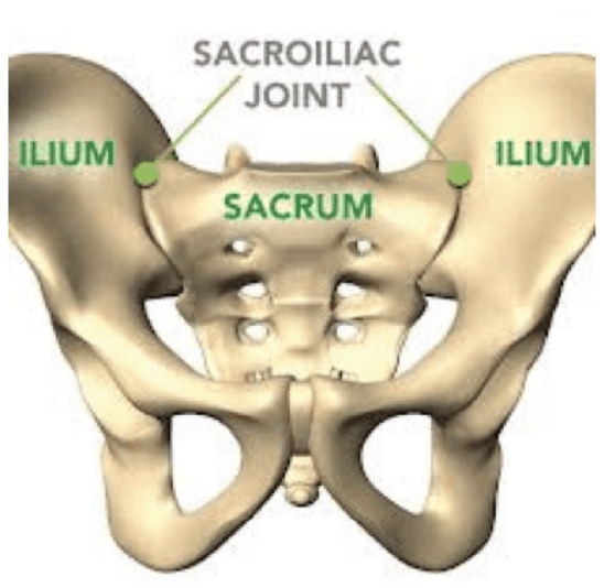
Sacroiliac (SI) Joint Fusion is a minimally invasive surgical procedure used to treat pain and instability caused by SI joint dysfunction. The sacroiliac joint connects the sacrum (the base of the spine) to the iliac bones (the pelvis). This joint plays a key role in transferring weight from the upper body to the legs and supports everyday movements like walking, bending, and lifting.
When the SI joint becomes unstable or inflamed—due to degeneration, trauma, or other causes—it can lead to chronic pain in the lower back, buttocks, or legs. SI joint fusion stabilizes the joint by placing an implant across it, helping to reduce pain and restore function.
Why Is a SI Joint Fusion Performed?
SI joint fusion is typically performed for patients with chronic SI joint pain or dysfunction that has not improved with non-surgical treatments. These may include:
- Physical therapy
- Chiropractic or osteopathic adjustments
- Acupuncture
- Anti-inflammatory medications
- SI joint injections or nerve blocks
If these conservative treatments fail to relieve pain or improve mobility, surgery may be considered as a long-term solution to stabilize the joint and improve quality of life.
How Do You Prepare for a SI Joint Fusion?
Preparation for SI joint fusion includes a full evaluation and imaging studies (such as X-rays, CT scans, or MRI) to confirm that the SI joint is the true source of pain. Your provider may also perform diagnostic injections to determine whether the joint responds to numbing medication.
Before surgery, you may be advised to:
- Stop certain medications (especially blood thinners)
- Fast the night before the procedure
- Quit smoking to improve healing
- Arrange for transportation and home support
- Prepare your home with mobility aids such as a walker or elevated seating
Your care team will provide detailed instructions specific to your case and recovery plan.
What Can You Expect During a SI Joint Fusion?
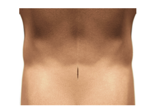
SI joint fusion is performed using minimally invasive techniques, which means less tissue disruption, smaller incisions, and faster healing.
Procedure Overview:
- You’ll receive general anesthesia or sedation for comfort.
- A small incision is made on the lower back, near the buttocks
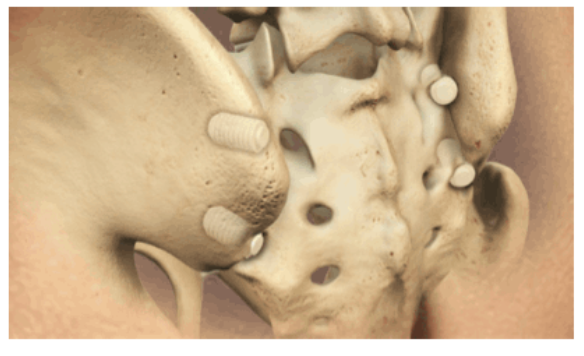
- Guide wires are inserted to help the surgeon position dilation tubes, which create a working channel through soft tissues without cutting through them.
- Through this channel, the surgeon drills and places a specialized implant across the SI joint to stop abnormal motion and allow the bones to fuse over time.
The procedure typically takes less than an hour, and patients are closely monitored during recovery. Dr. Kim uses computer navigation to maximize precision.
What Is the Recovery Like After a SI Joint Fusion?
Hospital Stay:
- Most patients go home same day following the procedure.
- A walker or cane may be used temporarily for support and to limit pressure on the joint during early healing.
Pain Management:
- Mild to moderate pain is common in the first few days, but it is usually well-managed with oral medications.
- Most patients notice gradual improvement in symptoms over several weeks.
Physical Therapy:
- Physical therapy usually begins 4 to 6 weeks after surgery and focuses on regaining strength, mobility, and balance.
- You’ll be guided through a personalized rehab program tailored to your level of activity and goals.
Returning to Work and Daily Life:
- Most patients return to work within 2 to 3 months, depending on the physical demands of their job.
- Lighter-duty tasks or remote work may be resumed sooner with clearance from your surgeon.
Your recovery timeline will be personalized based on your progress and overall health.
What Are the Potential Risks of a SI Joint Fusion?
SI joint fusion is a safe and effective procedure, but like any surgery, it does carry some risks. These may include:
- Infection
- Bleeding
- Nerve irritation
- Implant-related complications
- Delayed or incomplete fusion
- Persistent pain
Your surgeon will take every precaution to minimize risk and support a smooth recovery.
Are There Related Procedures to a SI Joint Fusion?
Yes. Depending on your specific condition and symptoms, related procedures may include:
- SI joint injections – for both diagnosis and temporary pain relief
- Radiofrequency ablation – to temporarily block nerve signals from the joint
- Physical therapy and manual therapy – as a first-line treatment or post-op support
- Spinal fusion – if pain is coming from multiple levels or adjacent joints in the spine
Your spine specialist will help determine whether SI joint fusion is the best treatment for your needs or if another option would be more appropriate.
Key Takeaways About SI Joint Fusion
- SI joint fusion is a minimally invasive surgery designed to stabilize the sacroiliac joint and relieve chronic pain.
- It is typically recommended after non-surgical treatments have failed.
- The procedure is performed through a small incision, using guidewires and implants to avoid major muscle or bone disruption.
- Patients are usually walking within a day or two, begin physical therapy within 4–6 weeks, and return to normal activities within a few months.
- The goal is to restore stability, reduce pain, and improve daily function.
Next Steps
If you’ve been struggling with persistent low back or pelvic pain and non-surgical treatments haven’t brought relief, SI joint fusion may be an effective next step. A thorough evaluation by a spine specialist can determine whether this minimally invasive solution is right for you.
